Wed, Oct 21, 2026


Volume 14, Issue 3 (9-2026)
J. Pediatr. Rev 2026, 14(3): 275-286 |
Back to browse issues page



Download citation:
BibTeX | RIS | EndNote | Medlars | ProCite | Reference Manager | RefWorks
Send citation to:


BibTeX | RIS | EndNote | Medlars | ProCite | Reference Manager | RefWorks
Send citation to:
Abdollahi G, Vahidi M R, Mousavi R S, Gashmard R, Mehboodi F, Mousavi S T. A Case Report of Thiamine-responsive Megaloblastic Anemia Misdiagnosis in a Male Child With Diabetic Ketoacidosis and Glucose-6-phosphate Dehydrogenase Deficiency. J. Pediatr. Rev 2026; 14 (3) :275-286
URL: http://jpr.mazums.ac.ir/article-1-864-en.html
URL: http://jpr.mazums.ac.ir/article-1-864-en.html
Gholamreza Abdollahi1 

 , Mohammad Reza Vahidi1 , Reyhaneh Sadat Mousavi1 , Roqayeh Gashmard2 , Farkhondeh Mehboodi3 , Seyedeh Tahereh Mousavi *4
1- Student Research Committee, Bushehr University of Medical Sciences, Bushehr, Iran.
2- Department of Pediatric Nursing, Faculty of Nursing and Midwifery, Bushehr University of Medical Sciences, Bushehr, Iran.
3- Department of Medical Surgical Nursing, Faculty of Nursing and Midwifery, Bushehr University of Medical Sciences, Bushehr, Iran.
4- Department of Pediatric Endocrinology, Faculty of Medicine, Bushehr University of Medical Sciences, Bushehr, Iran. ,taherehmousavi1979@gmail.com
2- Department of Pediatric Nursing, Faculty of Nursing and Midwifery, Bushehr University of Medical Sciences, Bushehr, Iran.
3- Department of Medical Surgical Nursing, Faculty of Nursing and Midwifery, Bushehr University of Medical Sciences, Bushehr, Iran.
4- Department of Pediatric Endocrinology, Faculty of Medicine, Bushehr University of Medical Sciences, Bushehr, Iran. ,
Keywords: Type 1 diabetes, Diabetic ketoacidosis (DKA), Favism, Anemia, Pediatrics, Thiamine-responsive megaloblastic anemia (TRMA)
Full-Text [PDF 434 kb]
(120 Downloads)
| Abstract (HTML) (363 Views)
Full-Text: (70 Views)
Introduction
Type 1 diabetes mellitus (T1DM) is one of the most common chronic diseases in childhood, resulting from autoimmune destruction of pancreatic β-cells, leading to reduced or absent insulin secretion. The classical triad of diabetes symptoms includes polyuria, polydipsia, and polyphagia [1, 2]. In addition to clinical findings, specialized laboratory tests also play an important role in confirming the diagnosis of T1DM. Among the most significant evaluations are diabetes‑related autoantibodies, such as anti–glutamic acid decarboxylase (GAD) and islet cell antibodies (ICA), which are recognized as key markers of pancreatic autoimmunity [3]. According to studies, diabetic ketoacidosis (DKA), a life-threatening medical emergency, may be the initial presentation in 15-80% of children with newly diagnosed T1DM [4-6]. During the treatment of DKA, blood glucose control in some patients, especially those with glucose-6-phosphate dehydrogenase (G6PD) deficiency, can predispose to hemolytic anemia. G6PD deficiency is the most common enzymatic disorder in humans, which increases red blood cell susceptibility to oxidative stress and may lead to premature hemolysis in situations such as DKA correction [4, 7]. However, most individuals with G6PD deficiency remain asymptomatic until exposed to triggering factors such as fava bean (Broad Bean) ingestion, certain medications, severe infections, renal failure, or DKA [8].
On the other hand, the co-occurrence of macrocytic anemia and diabetes may raise suspicion for thiamine-responsive megaloblastic anemia (TRMA), a rare genetic disorder caused by mutations in the SLC19A2 gene and characterized by the classic triad of megaloblastic anemia, diabetes mellitus, and progressive sensorineural hearing loss [9, 10]. This diagnosis may appear more plausible when a patient enters the honeymoon phase of T1DM, during which insulin requirements decrease. This phenomenon might be misinterpreted as a favorable response to thiamine therapy [9, 11]. Discontinuation of insulin therapy in such cases may lead to recurrence of DKA symptoms, including polyuria, polydipsia, weight loss, fatigue, and respiratory distress [2, 12]. In this study, we report a rare case of a child with newly diagnosed T1DM presenting with DKA who developed anemia due to G6PD deficiency-induced hemolysis but was initially misdiagnosed with TRMA and managed accordingly, eventually leading to the recurrence of DKA symptoms and re-hospitalization.
Case Presentation
The case was a 5-year, 2-month, and 9-day-old boy who was admitted to a hospital in Bushehr, south of Iran, with symptoms of fever, Kussmaul respirations, weakness, lethargy, fatigue, anorexia, polydipsia, polyuria, nausea, vomiting, abdominal pain, decreased level of consciousness, and moderate dehydration. Approximately five days before admission, he had developed fever and cough in the context of an upper respiratory tract infection, during which he was twice evaluated by physicians and treated with intravenous fluids and medications. However, symptoms such as anorexia, polydipsia, and polyuria had been present a few weeks earlier. At the time of admission, the child was toxic and presented with respiratory distress. On lung auscultation, wheezing and crackles were heard, particularly in the right lung.
Patient background
He was the second child in the family, with a birth weight of approximately 3800 grams. His growth and developmental indicators were within normal limits. There was no prior history of hospitalization, medication use, or surgical procedures. In the neonatal period, he had prolonged jaundice, which, although potentially indicative of G6PD deficiency (favism), had not been investigated at the time. His older sibling (16 years old) was healthy with only a history of febrile seizures. Their parents were distantly related. There was no family history of diabetes mellitus, but maternal relatives had a history of thyroid disorders.
First hospital admission
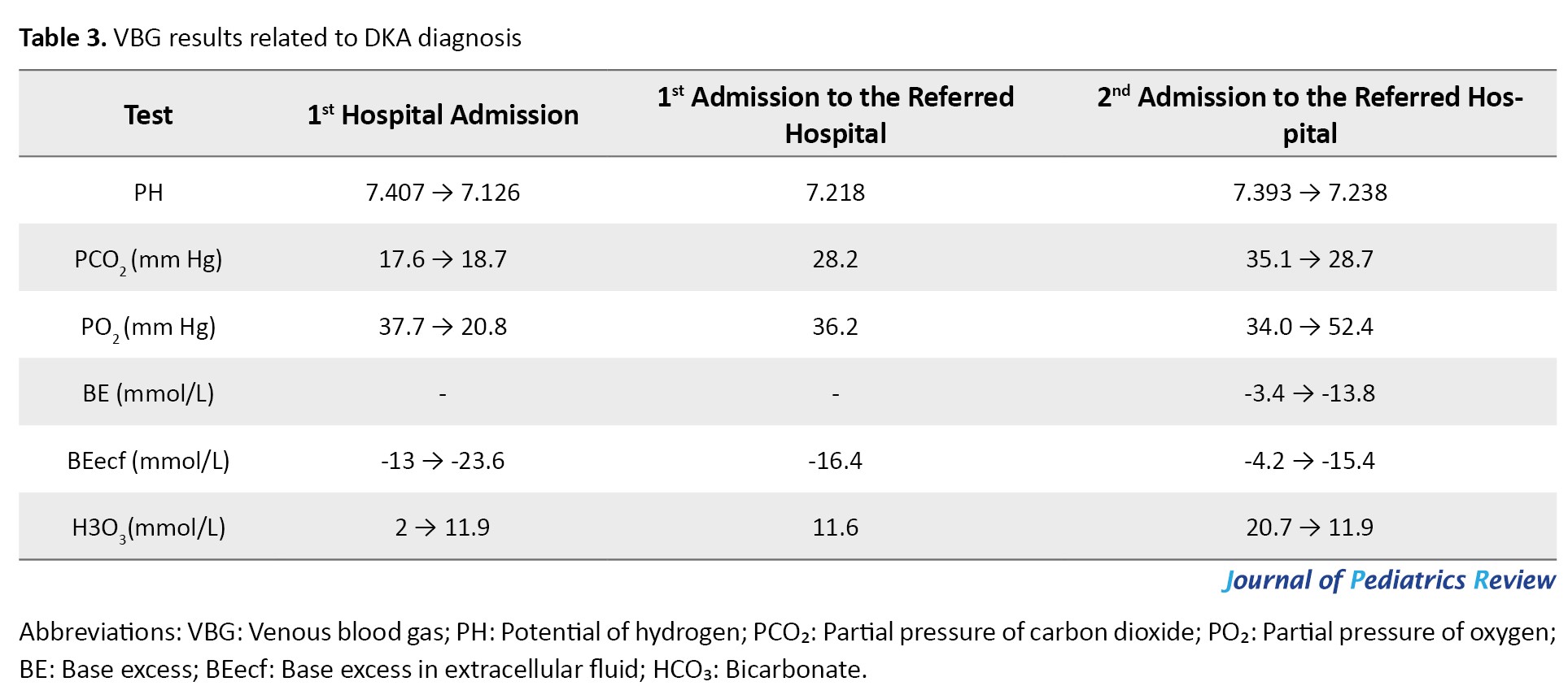
Upon admission to the hospital, the patient was diagnosed with DKA based on clinical findings and laboratory results (Tables 1, 2 and 3).
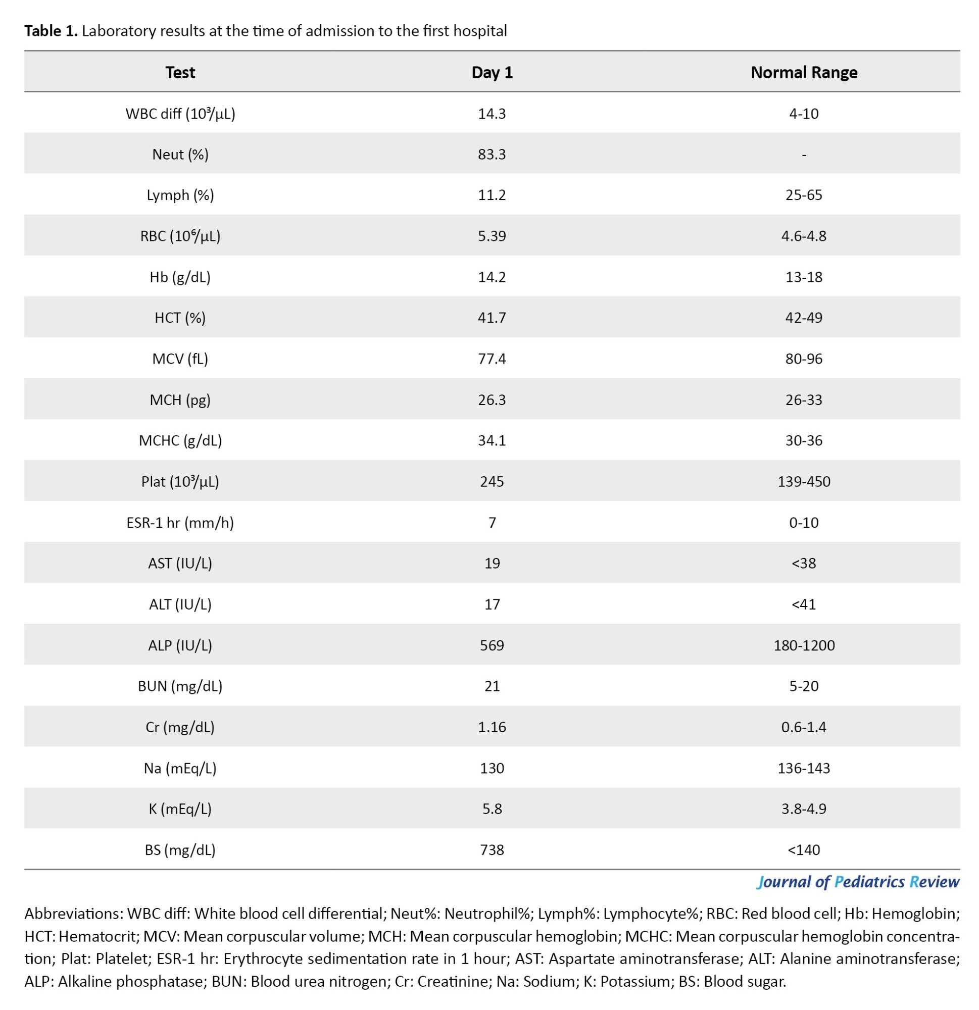
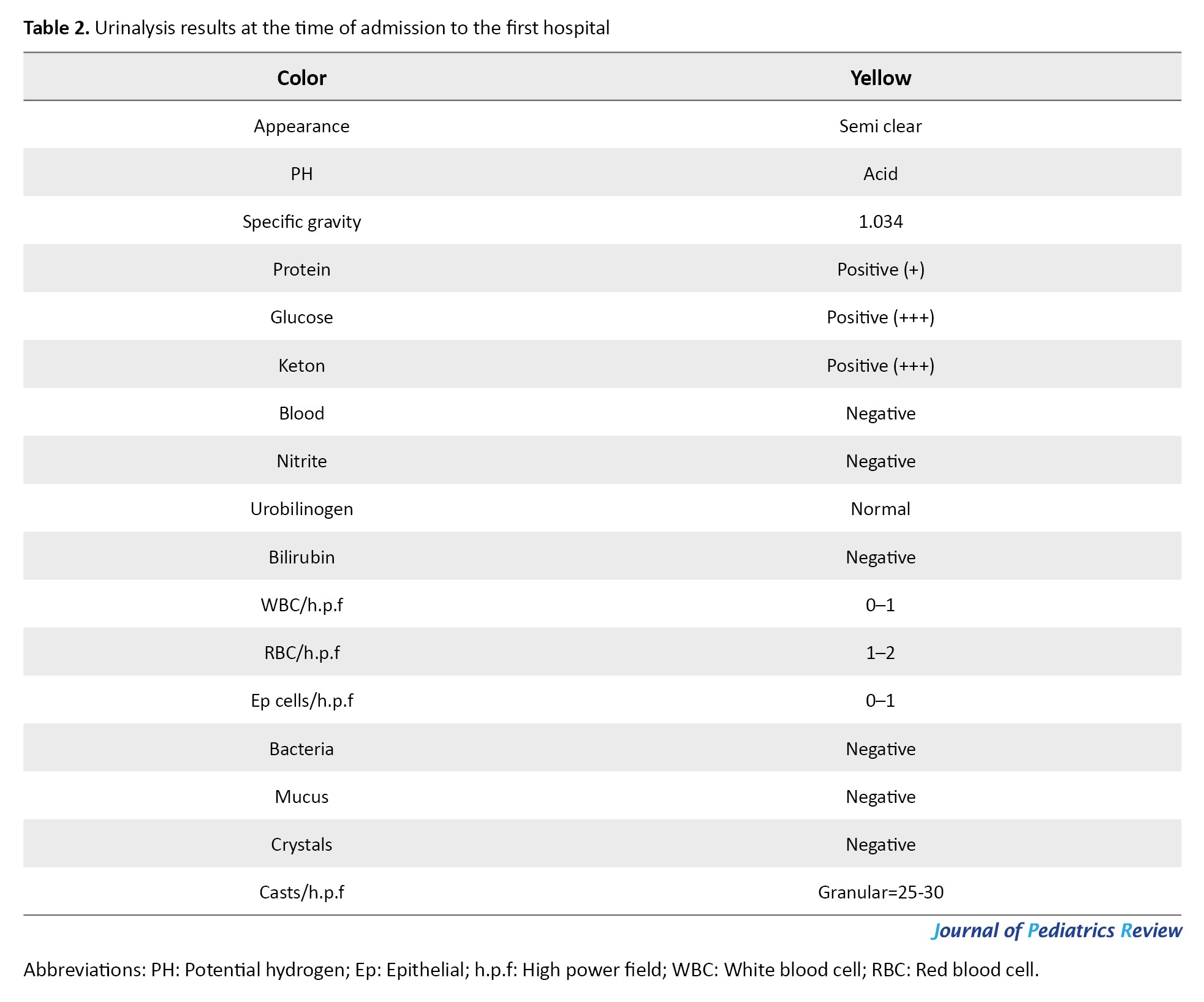
Treatment was promptly initiated, including fluid therapy that started with a bolus of normal saline (20 cc/kg over the first hour), followed by half-normal saline (0.45% NaCl), calculated based on maintenance requirements and a 10% dehydration correction. Regular insulin infusion was started at a dose of 0.1 unit/kg/hr. Supportive care and monitoring of electrolytes, serum composition, and insulin dose adjustments were made according to laboratory parameters and clinical evolution (Table 1). Given the history of fever, intravenous antibiotic therapy (ceftriaxone at 75 mg/kg/day divided into two doses) was administered. Due to a decreased level of consciousness at the 18th hour of hospitalization, cerebral edema was suspected, and the patient received two doses of intravenous mannitol at 1 g/kg. On the second day of hospitalization, he was referred to another hospital in Bushehr.
First admission to the referred hospital
On the second day of treatment, the patient was transferred to the referred hospital. Upon admission, the patient’s vital signs were as follows: Blood pressure: 90/60 mm Hg, pulse rate: 117 bpm, respiratory rate: 28 breaths/min, temperature: 38.5 °C, blood sugar: 263 mg/dL, pH: 7.218, HCO₃⁻: 11.6 mmol/L, PCO₂: 28.2 mm Hg. The patient was alert, with a body weight of 14 kg at arrival.
In the emergency department, clinical and laboratory findings (Table 4) confirmed a diagnosis of new-onset T1DM.
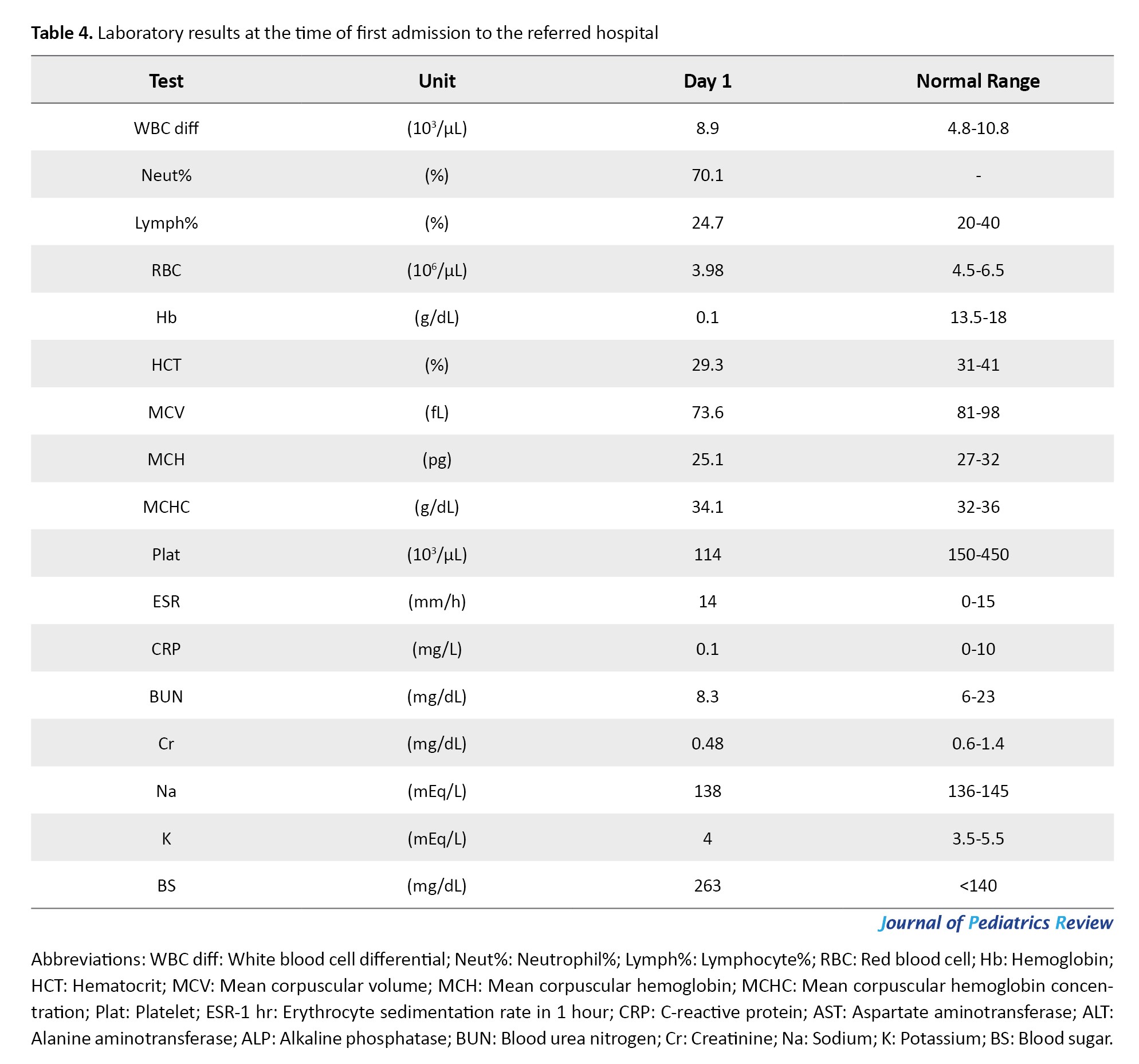
Given the fever, abnormal lung examination findings, and chest radiography, pneumonia was also diagnosed. Accordingly, in addition to intravenous ceftriaxone, clindamycin was initiated, along with supportive treatments including normal saline infusion, Apotel, 3% NaCl nebulization, and Ventolin nebulization. DKA management was continued until resolution of the acute phase. Upon completion of DKA treatment, subcutaneous insulin therapy with Novorapid and Lantus was initiated. During hospitalization, the patient received ongoing supportive care, including nebulized therapy, intravenous fluids, and antibiotic therapy. Capillary blood glucose was monitored before and two hours after each meal, and insulin doses were adjusted accordingly to maintain glycemic control. With clinical improvement and resolution of pneumonia, clindamycin was discontinued on day 5, and ceftriaxone was stopped on day 6 of hospitalization. Finally, on day 6, the patient was discharged in good overall condition, with outpatient follow-up. Before discharge, the patient’s parents received comprehensive education on insulin administration and home blood glucose monitoring.
Diagnosis of thiamine-responsive megaloblastic anemia after discharge
Approximately one week after discharge, the patient was referred to the hospital for follow-up of blood glucose levels. Given the acceptable glycemic profile and several episodes of hypoglycemia in the preceding days, insulin dosage was reduced under the assumption that the patient had entered the honeymoon phase of T1DM. During the clinical evaluation, the child appeared significantly weak, pale, and lethargic, with impaired mobility due to profound fatigue. Consequently, a new set of laboratory tests was ordered (Table 5).
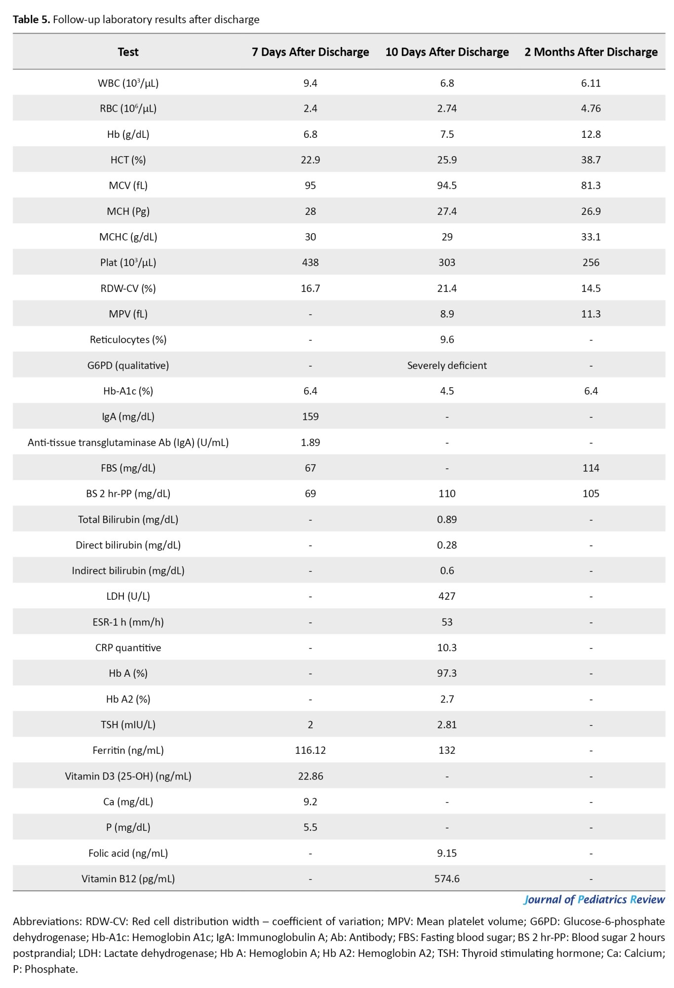
The results showed a hemoglobin level of 8.6 g/dL, red blood cell (RBC) count of 4.2 million/µL, and mean corpuscular volume (MCV) of 95 fL. Because of the anemia, a hematology consultation was requested for further diagnostic evaluation. According to the results of the requested laboratory tests (Table 5) and considering the presence of macrocytic anemia concurrently with diabetes, the possibility of TRMA was considered as the initial diagnosis. Accordingly, treatment with oral thiamine (100 mg twice daily) and folic acid (5 mg once daily) was initiated. At the same time, insulin therapy was discontinued, coinciding with the beginning of the honeymoon phase of T1DM, a period during which insulin requirements temporarily decline, and normoglycemia may be maintained without insulin. Reassured by the new diagnosis, the child’s family decided to discontinue insulin therapy and cease diabetes-related follow-up. This continued until the child exited the honeymoon phase, at which point hyperglycemia recurred, and he was readmitted to the referred hospital with a second episode of DKA.
Second admission to the referred hospital
Three months after the initial diagnosis of T1DM, the patient was readmitted to the referred hospital with symptoms of fatigue, lethargy, constipation, polyuria, and polydipsia. At the time of admission, his vital signs were as follows: Blood pressure: 95/55 mm Hg, pulse rate: 103 bpm, respiratory rate: 20 breaths/min, temperature: 37.2 °C. A home blood glucose measurement prior to admission was 320 mg/dL. The patient was alert and weighed 13.5 kg upon arrival. Based on laboratory findings (Hb: 12.1 g/dL, blood sugar: 352 mg/dL, pH: 7.238, HCO₃⁻: 11.9 mmol/L, PCO₂: 28.7 mm Hg), a second diagnosis of DKA was established (Tables 3, 6 and 7), and the standard treatment for DKA was initiated.
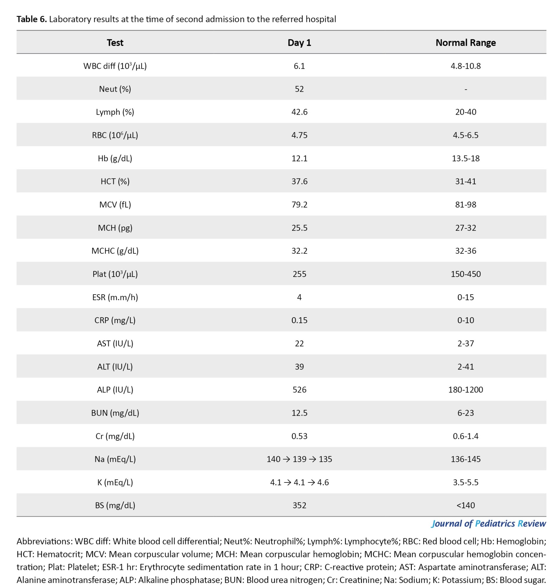
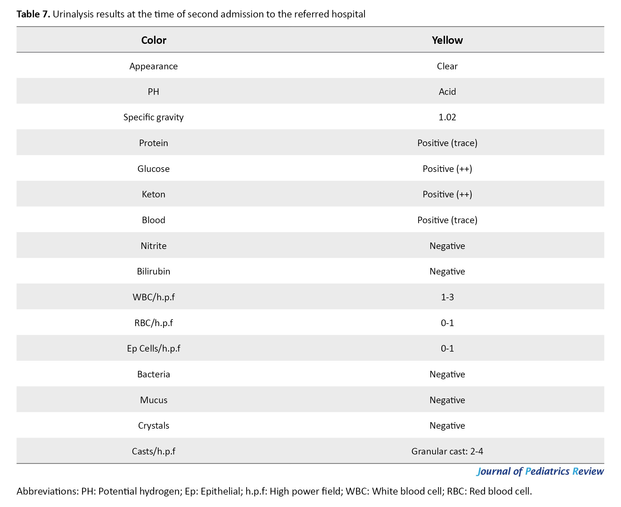
During hospitalization and further investigations, it was determined that the previous diagnosis of TRMA was incorrect, and the patient’s anemia was actually related to G6PD deficiency. Consequently, treatment with thiamine was discontinued, and insulin therapy was restarted. The patient was discharged after clinical stabilization, with an appropriate diet and insulin treatment plan. At discharge, subcutaneous insulin therapy with Lantus and Novorapid was reinitiated, thiamine tablets were discontinued, and folic acid (5 mg once daily) was continued. The family was advised to strictly avoid fava beans and foods high in vitamin C, to limit prolonged physical activity, and to ensure adequate intake of iron, folic acid, and foods rich in vitamin E and selenium. They were also instructed to avoid medications such as aspirin and ciprofloxacin. Most importantly, the family was strongly advised to inform the healthcare team of the patient’s G6PD deficiency (favism) before initiating any medical intervention.
Follow-up
The latest follow-up was conducted when the patient was 5 years and 6 months old, approximately four months after the initial diagnosis of T1DM. At this time, the patient had appropriate weight gain, reaching 18 kilograms. Blood glucose levels were well controlled, and HbA1c was within the acceptable range. The patient showed no signs of anemia, and the CBC revealed normal parameters related to anemia, as shown in Table 8.
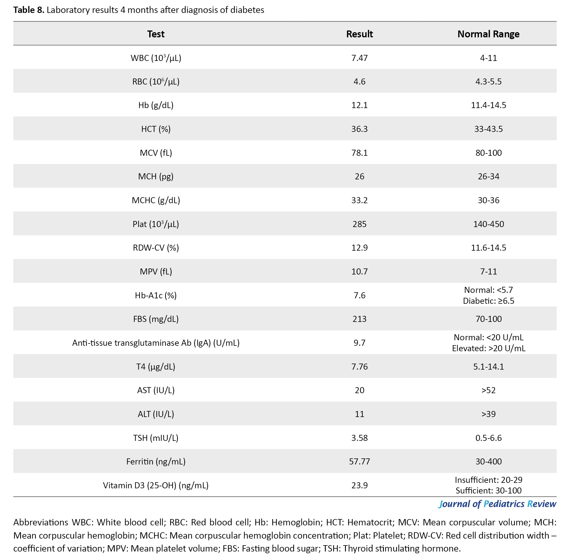
Currently, the patient is in stable condition and continues to receive regular insulin therapy.
Discussion
In this study, we presented a 5-year-old child with T1DM and G6PD deficiency who developed DKA and hemolysis during treatment. Insulin-dependent T1DM is the most common form of diabetes in children, and up to 40% of cases may initially present with DKA. This condition often results from a lack of attention to classic diabetes symptoms [2, 5, 6, 13, 14]. According to the European Society for Pediatric Endocrinology and the Lawson Wilkins Pediatric Endocrine Society (ESPE/LWPES) consensus statement, hyperglycemia (blood sugar ≥200 mg/dL) in the presence of metabolic acidosis (pH <7.3, HCO3 <18) and moderate to severe ketonemia or ketonuria are biochemical criteria for diagnosing DKA. Clinical manifestations include dehydration, Kussmaul respiration, weakness, lethargy, decreased consciousness level, and in severe cases, coma. Predisposing factors include delayed diagnosis, resulting in late initiation of treatment, as well as the presence of concurrent infections. A history of recent febrile illness or common cold has been reported to be associated with approximately a tenfold increase in the number of children with T1DM and DKA at the time of diagnosis [15-17]. In our patient, both predisposing factors were relevant, despite the presence of polyuria and polydipsia several weeks earlier. Due to family neglect and delayed diagnosis at outpatient centers, the child presented to the emergency department with dehydration, decreased consciousness, blood glucose of 738 mg/dL, pH=7.12, and HCO3 < 5 mmol/L, leading to hospitalization.
G6PD is an X-linked enzyme that protects RBCs from oxidative stress. Deficiency of this enzyme in the presence of oxidative triggers such as certain foods (e.g. fava beans), drugs (antimalarials, anticonvulsants, vitamin C), infections, and metabolic stresses (such as DKA) can result in acute hemolytic episodes. Clinical manifestations of hemolysis due to this deficiency may include weakness, pallor, lethargy, and urine discoloration [12, 18, 19]. Furthermore, in the early stages of G6PD deficiency-related hemolysis, thrombocytosis or mild microcytic anemia may be observed; however, during the recovery phase, reticulocytosis leads to increased MCV and a macrocytic anemia picture [19-22]. In our patient, the two main hemolytic triggers were acute pulmonary infection (pneumonia) and DKA. However, during hospitalization, hemolytic symptoms such as urine discoloration were not clinically significant, and urinalysis showed no clear findings. The severity of anemia at admission (hemoglobin 10 g/dL) also did not warrant urgent evaluation. Therefore, the treatment team focused on managing pneumonia, adjusting insulin doses, controlling blood glucose, and educating the family on post-discharge diabetic care. Given the high prevalence of G6PD deficiency in Iran (10–14% according to the World Health Organization) and in its southern cities like Bushehr, this diagnosis should be considered in all cases of symptomatic acute anemia, especially when accompanied by urine discoloration, although our patient did not have classic clinical features of G6PD deficiency [23]. Therefore, further investigations were conducted after his discharge to determine the cause of anemia.
Since the simultaneous occurrence of macrocytic anemia and diabetes can raise suspicion for TRMA, this diagnosis was considered based on the laboratory results (Hb=6.8 and MCV=95). TRMA is a rare genetic disorder in which thiamine administration can improve macrocytic anemia and reduce insulin requirements, although it may not significantly affect other manifestations of the syndrome, such as sensorineural hearing loss. Therefore, audiological evaluation with audiometry is considered an important part of the diagnostic workup for patients suspected of TRMA [9, 10, 24]. However, in our case, audiometric evaluation was not performed early. Nevertheless, thiamine was administered at a dose of 100 mg twice daily, and a partial improvement in clinical status was observed, which was initially interpreted as a response to thiamine treatment. This continued until the patient exited the “honeymoon” phase, with a subsequent rise in blood glucose and a second DKA episode requiring hospital readmission.
It became clear that although the healthcare team requested initial investigations for anemia, the diagnosis of TRMA was premature. It would have been more appropriate to consider all differential diagnoses one by one and wait for the results of initial tests. A more thorough history-taking, along with a detailed evaluation of the patient’s symptoms and the trend of CBCs before and during hospitalization, should have been conducted. The possibility of the “honeymoon” phase as the cause of transient hypoglycemia should have been considered from the outset. Even if clinical doubt persisted, a gradual tapering of the insulin dose would have been more appropriate than abrupt discontinuation, especially given the family’s reluctance to accept a permanent diabetes diagnosis. Therefore, other potential causes of macrocytic anemia in a diabetic child, including vitamin B12 and folate deficiency, hypothyroidism, malabsorption syndromes such as celiac disease, and liver disease, were evaluated. Normal thyroid function tests (TSH: 2.81), liver enzymes (AST: 19, ALP: 569, ALT: 17), and negative gluten sensitivity excluded these conditions [25, 26].
Finally, considering that reticulocytosis can cause increased MCV alongside anemia, and that various hemolytic anemias, including G6PD deficiency-related hemolysis, can present similarly, it was determined that the diagnosis of TRMA was made prematurely, and in fact, the anemia resulted from RBC lysis due to G6PD deficiency (favism) in the context of DKA and infection.
Conclusion
The coexistence of G6PD deficiency and the possible honeymoon phase of T1DM led to misdiagnosis of TRMA in a male child. This highlights the importance of step-by-step differential diagnosis, accurate interpretation of laboratory results, and avoiding hasty clinical decisions. Moreover, even when the diagnosis of TRMA is accurate, insulin therapy must be discontinued gradually and with close blood glucose monitoring. This is crucial, as in some TRMA cases, diabetes does not respond to thiamine and insulin therapy remains necessary. Therefore, proper family education and emphasis on regular follow-up play a vital role in preventing complications.
Ethical Considerations
Compliance with ethical guidelines
Ethics approval for this case report was obtained from the Ethics Committee of Bushehr University of Medical Sciences, Bushehr, Iran (Code: IR.BPUMS.REC.1404.407). Written informed consent was obtained from the patient’s legal guardian.
Funding
This research did not receive any specific grant from funding agencies in the public, commercial, or not-for profit sectors.
Authors contributions
Supervision: Seyedeh Tahereh Mousavi, Roqayeh Gashmard, and Farkhondeh Mehboodi; Data collection: Mohammad Reza Vahidi, Reyhaneh Sadat Mousavi, and Gholamreza Abdollahi; Clinical case management and clinical interpretation: Seyedeh Tahereh Mousavi; Writing the original draft: Gholamreza Abdollahi, Reyhaneh Sadat Mousavi, Mohammad Reza Vahidi, and Seyedeh Tahereh Mousavi; Final approval: All authors.
Conflicts of interest
The authors declared no conflict of interest.
Acknowledgements
The authors would like to thank the Student Research Committee and the Vice-Chancellor for Research and Technology of Bushehr University of Medical Sciences, the Clinical Research Development Unit and the management of the Persian Gulf Martyrs Hospital in Bushehr, as well as the patient and his family for their time and cooperation throughout this study.
Type 1 diabetes mellitus (T1DM) is one of the most common chronic diseases in childhood, resulting from autoimmune destruction of pancreatic β-cells, leading to reduced or absent insulin secretion. The classical triad of diabetes symptoms includes polyuria, polydipsia, and polyphagia [1, 2]. In addition to clinical findings, specialized laboratory tests also play an important role in confirming the diagnosis of T1DM. Among the most significant evaluations are diabetes‑related autoantibodies, such as anti–glutamic acid decarboxylase (GAD) and islet cell antibodies (ICA), which are recognized as key markers of pancreatic autoimmunity [3]. According to studies, diabetic ketoacidosis (DKA), a life-threatening medical emergency, may be the initial presentation in 15-80% of children with newly diagnosed T1DM [4-6]. During the treatment of DKA, blood glucose control in some patients, especially those with glucose-6-phosphate dehydrogenase (G6PD) deficiency, can predispose to hemolytic anemia. G6PD deficiency is the most common enzymatic disorder in humans, which increases red blood cell susceptibility to oxidative stress and may lead to premature hemolysis in situations such as DKA correction [4, 7]. However, most individuals with G6PD deficiency remain asymptomatic until exposed to triggering factors such as fava bean (Broad Bean) ingestion, certain medications, severe infections, renal failure, or DKA [8].
On the other hand, the co-occurrence of macrocytic anemia and diabetes may raise suspicion for thiamine-responsive megaloblastic anemia (TRMA), a rare genetic disorder caused by mutations in the SLC19A2 gene and characterized by the classic triad of megaloblastic anemia, diabetes mellitus, and progressive sensorineural hearing loss [9, 10]. This diagnosis may appear more plausible when a patient enters the honeymoon phase of T1DM, during which insulin requirements decrease. This phenomenon might be misinterpreted as a favorable response to thiamine therapy [9, 11]. Discontinuation of insulin therapy in such cases may lead to recurrence of DKA symptoms, including polyuria, polydipsia, weight loss, fatigue, and respiratory distress [2, 12]. In this study, we report a rare case of a child with newly diagnosed T1DM presenting with DKA who developed anemia due to G6PD deficiency-induced hemolysis but was initially misdiagnosed with TRMA and managed accordingly, eventually leading to the recurrence of DKA symptoms and re-hospitalization.
Case Presentation
The case was a 5-year, 2-month, and 9-day-old boy who was admitted to a hospital in Bushehr, south of Iran, with symptoms of fever, Kussmaul respirations, weakness, lethargy, fatigue, anorexia, polydipsia, polyuria, nausea, vomiting, abdominal pain, decreased level of consciousness, and moderate dehydration. Approximately five days before admission, he had developed fever and cough in the context of an upper respiratory tract infection, during which he was twice evaluated by physicians and treated with intravenous fluids and medications. However, symptoms such as anorexia, polydipsia, and polyuria had been present a few weeks earlier. At the time of admission, the child was toxic and presented with respiratory distress. On lung auscultation, wheezing and crackles were heard, particularly in the right lung.
Patient background
He was the second child in the family, with a birth weight of approximately 3800 grams. His growth and developmental indicators were within normal limits. There was no prior history of hospitalization, medication use, or surgical procedures. In the neonatal period, he had prolonged jaundice, which, although potentially indicative of G6PD deficiency (favism), had not been investigated at the time. His older sibling (16 years old) was healthy with only a history of febrile seizures. Their parents were distantly related. There was no family history of diabetes mellitus, but maternal relatives had a history of thyroid disorders.
First hospital admission
Upon admission to the hospital, the patient was diagnosed with DKA based on clinical findings and laboratory results (Tables 1, 2 and 3).
Treatment was promptly initiated, including fluid therapy that started with a bolus of normal saline (20 cc/kg over the first hour), followed by half-normal saline (0.45% NaCl), calculated based on maintenance requirements and a 10% dehydration correction. Regular insulin infusion was started at a dose of 0.1 unit/kg/hr. Supportive care and monitoring of electrolytes, serum composition, and insulin dose adjustments were made according to laboratory parameters and clinical evolution (Table 1). Given the history of fever, intravenous antibiotic therapy (ceftriaxone at 75 mg/kg/day divided into two doses) was administered. Due to a decreased level of consciousness at the 18th hour of hospitalization, cerebral edema was suspected, and the patient received two doses of intravenous mannitol at 1 g/kg. On the second day of hospitalization, he was referred to another hospital in Bushehr.
First admission to the referred hospital
On the second day of treatment, the patient was transferred to the referred hospital. Upon admission, the patient’s vital signs were as follows: Blood pressure: 90/60 mm Hg, pulse rate: 117 bpm, respiratory rate: 28 breaths/min, temperature: 38.5 °C, blood sugar: 263 mg/dL, pH: 7.218, HCO₃⁻: 11.6 mmol/L, PCO₂: 28.2 mm Hg. The patient was alert, with a body weight of 14 kg at arrival.
In the emergency department, clinical and laboratory findings (Table 4) confirmed a diagnosis of new-onset T1DM.
Given the fever, abnormal lung examination findings, and chest radiography, pneumonia was also diagnosed. Accordingly, in addition to intravenous ceftriaxone, clindamycin was initiated, along with supportive treatments including normal saline infusion, Apotel, 3% NaCl nebulization, and Ventolin nebulization. DKA management was continued until resolution of the acute phase. Upon completion of DKA treatment, subcutaneous insulin therapy with Novorapid and Lantus was initiated. During hospitalization, the patient received ongoing supportive care, including nebulized therapy, intravenous fluids, and antibiotic therapy. Capillary blood glucose was monitored before and two hours after each meal, and insulin doses were adjusted accordingly to maintain glycemic control. With clinical improvement and resolution of pneumonia, clindamycin was discontinued on day 5, and ceftriaxone was stopped on day 6 of hospitalization. Finally, on day 6, the patient was discharged in good overall condition, with outpatient follow-up. Before discharge, the patient’s parents received comprehensive education on insulin administration and home blood glucose monitoring.
Diagnosis of thiamine-responsive megaloblastic anemia after discharge
Approximately one week after discharge, the patient was referred to the hospital for follow-up of blood glucose levels. Given the acceptable glycemic profile and several episodes of hypoglycemia in the preceding days, insulin dosage was reduced under the assumption that the patient had entered the honeymoon phase of T1DM. During the clinical evaluation, the child appeared significantly weak, pale, and lethargic, with impaired mobility due to profound fatigue. Consequently, a new set of laboratory tests was ordered (Table 5).
The results showed a hemoglobin level of 8.6 g/dL, red blood cell (RBC) count of 4.2 million/µL, and mean corpuscular volume (MCV) of 95 fL. Because of the anemia, a hematology consultation was requested for further diagnostic evaluation. According to the results of the requested laboratory tests (Table 5) and considering the presence of macrocytic anemia concurrently with diabetes, the possibility of TRMA was considered as the initial diagnosis. Accordingly, treatment with oral thiamine (100 mg twice daily) and folic acid (5 mg once daily) was initiated. At the same time, insulin therapy was discontinued, coinciding with the beginning of the honeymoon phase of T1DM, a period during which insulin requirements temporarily decline, and normoglycemia may be maintained without insulin. Reassured by the new diagnosis, the child’s family decided to discontinue insulin therapy and cease diabetes-related follow-up. This continued until the child exited the honeymoon phase, at which point hyperglycemia recurred, and he was readmitted to the referred hospital with a second episode of DKA.
Second admission to the referred hospital
Three months after the initial diagnosis of T1DM, the patient was readmitted to the referred hospital with symptoms of fatigue, lethargy, constipation, polyuria, and polydipsia. At the time of admission, his vital signs were as follows: Blood pressure: 95/55 mm Hg, pulse rate: 103 bpm, respiratory rate: 20 breaths/min, temperature: 37.2 °C. A home blood glucose measurement prior to admission was 320 mg/dL. The patient was alert and weighed 13.5 kg upon arrival. Based on laboratory findings (Hb: 12.1 g/dL, blood sugar: 352 mg/dL, pH: 7.238, HCO₃⁻: 11.9 mmol/L, PCO₂: 28.7 mm Hg), a second diagnosis of DKA was established (Tables 3, 6 and 7), and the standard treatment for DKA was initiated.
During hospitalization and further investigations, it was determined that the previous diagnosis of TRMA was incorrect, and the patient’s anemia was actually related to G6PD deficiency. Consequently, treatment with thiamine was discontinued, and insulin therapy was restarted. The patient was discharged after clinical stabilization, with an appropriate diet and insulin treatment plan. At discharge, subcutaneous insulin therapy with Lantus and Novorapid was reinitiated, thiamine tablets were discontinued, and folic acid (5 mg once daily) was continued. The family was advised to strictly avoid fava beans and foods high in vitamin C, to limit prolonged physical activity, and to ensure adequate intake of iron, folic acid, and foods rich in vitamin E and selenium. They were also instructed to avoid medications such as aspirin and ciprofloxacin. Most importantly, the family was strongly advised to inform the healthcare team of the patient’s G6PD deficiency (favism) before initiating any medical intervention.
Follow-up
The latest follow-up was conducted when the patient was 5 years and 6 months old, approximately four months after the initial diagnosis of T1DM. At this time, the patient had appropriate weight gain, reaching 18 kilograms. Blood glucose levels were well controlled, and HbA1c was within the acceptable range. The patient showed no signs of anemia, and the CBC revealed normal parameters related to anemia, as shown in Table 8.
Currently, the patient is in stable condition and continues to receive regular insulin therapy.
Discussion
In this study, we presented a 5-year-old child with T1DM and G6PD deficiency who developed DKA and hemolysis during treatment. Insulin-dependent T1DM is the most common form of diabetes in children, and up to 40% of cases may initially present with DKA. This condition often results from a lack of attention to classic diabetes symptoms [2, 5, 6, 13, 14]. According to the European Society for Pediatric Endocrinology and the Lawson Wilkins Pediatric Endocrine Society (ESPE/LWPES) consensus statement, hyperglycemia (blood sugar ≥200 mg/dL) in the presence of metabolic acidosis (pH <7.3, HCO3 <18) and moderate to severe ketonemia or ketonuria are biochemical criteria for diagnosing DKA. Clinical manifestations include dehydration, Kussmaul respiration, weakness, lethargy, decreased consciousness level, and in severe cases, coma. Predisposing factors include delayed diagnosis, resulting in late initiation of treatment, as well as the presence of concurrent infections. A history of recent febrile illness or common cold has been reported to be associated with approximately a tenfold increase in the number of children with T1DM and DKA at the time of diagnosis [15-17]. In our patient, both predisposing factors were relevant, despite the presence of polyuria and polydipsia several weeks earlier. Due to family neglect and delayed diagnosis at outpatient centers, the child presented to the emergency department with dehydration, decreased consciousness, blood glucose of 738 mg/dL, pH=7.12, and HCO3 < 5 mmol/L, leading to hospitalization.
G6PD is an X-linked enzyme that protects RBCs from oxidative stress. Deficiency of this enzyme in the presence of oxidative triggers such as certain foods (e.g. fava beans), drugs (antimalarials, anticonvulsants, vitamin C), infections, and metabolic stresses (such as DKA) can result in acute hemolytic episodes. Clinical manifestations of hemolysis due to this deficiency may include weakness, pallor, lethargy, and urine discoloration [12, 18, 19]. Furthermore, in the early stages of G6PD deficiency-related hemolysis, thrombocytosis or mild microcytic anemia may be observed; however, during the recovery phase, reticulocytosis leads to increased MCV and a macrocytic anemia picture [19-22]. In our patient, the two main hemolytic triggers were acute pulmonary infection (pneumonia) and DKA. However, during hospitalization, hemolytic symptoms such as urine discoloration were not clinically significant, and urinalysis showed no clear findings. The severity of anemia at admission (hemoglobin 10 g/dL) also did not warrant urgent evaluation. Therefore, the treatment team focused on managing pneumonia, adjusting insulin doses, controlling blood glucose, and educating the family on post-discharge diabetic care. Given the high prevalence of G6PD deficiency in Iran (10–14% according to the World Health Organization) and in its southern cities like Bushehr, this diagnosis should be considered in all cases of symptomatic acute anemia, especially when accompanied by urine discoloration, although our patient did not have classic clinical features of G6PD deficiency [23]. Therefore, further investigations were conducted after his discharge to determine the cause of anemia.
Since the simultaneous occurrence of macrocytic anemia and diabetes can raise suspicion for TRMA, this diagnosis was considered based on the laboratory results (Hb=6.8 and MCV=95). TRMA is a rare genetic disorder in which thiamine administration can improve macrocytic anemia and reduce insulin requirements, although it may not significantly affect other manifestations of the syndrome, such as sensorineural hearing loss. Therefore, audiological evaluation with audiometry is considered an important part of the diagnostic workup for patients suspected of TRMA [9, 10, 24]. However, in our case, audiometric evaluation was not performed early. Nevertheless, thiamine was administered at a dose of 100 mg twice daily, and a partial improvement in clinical status was observed, which was initially interpreted as a response to thiamine treatment. This continued until the patient exited the “honeymoon” phase, with a subsequent rise in blood glucose and a second DKA episode requiring hospital readmission.
It became clear that although the healthcare team requested initial investigations for anemia, the diagnosis of TRMA was premature. It would have been more appropriate to consider all differential diagnoses one by one and wait for the results of initial tests. A more thorough history-taking, along with a detailed evaluation of the patient’s symptoms and the trend of CBCs before and during hospitalization, should have been conducted. The possibility of the “honeymoon” phase as the cause of transient hypoglycemia should have been considered from the outset. Even if clinical doubt persisted, a gradual tapering of the insulin dose would have been more appropriate than abrupt discontinuation, especially given the family’s reluctance to accept a permanent diabetes diagnosis. Therefore, other potential causes of macrocytic anemia in a diabetic child, including vitamin B12 and folate deficiency, hypothyroidism, malabsorption syndromes such as celiac disease, and liver disease, were evaluated. Normal thyroid function tests (TSH: 2.81), liver enzymes (AST: 19, ALP: 569, ALT: 17), and negative gluten sensitivity excluded these conditions [25, 26].
Finally, considering that reticulocytosis can cause increased MCV alongside anemia, and that various hemolytic anemias, including G6PD deficiency-related hemolysis, can present similarly, it was determined that the diagnosis of TRMA was made prematurely, and in fact, the anemia resulted from RBC lysis due to G6PD deficiency (favism) in the context of DKA and infection.
Conclusion
The coexistence of G6PD deficiency and the possible honeymoon phase of T1DM led to misdiagnosis of TRMA in a male child. This highlights the importance of step-by-step differential diagnosis, accurate interpretation of laboratory results, and avoiding hasty clinical decisions. Moreover, even when the diagnosis of TRMA is accurate, insulin therapy must be discontinued gradually and with close blood glucose monitoring. This is crucial, as in some TRMA cases, diabetes does not respond to thiamine and insulin therapy remains necessary. Therefore, proper family education and emphasis on regular follow-up play a vital role in preventing complications.
Ethical Considerations
Compliance with ethical guidelines
Ethics approval for this case report was obtained from the Ethics Committee of Bushehr University of Medical Sciences, Bushehr, Iran (Code: IR.BPUMS.REC.1404.407). Written informed consent was obtained from the patient’s legal guardian.
Funding
This research did not receive any specific grant from funding agencies in the public, commercial, or not-for profit sectors.
Authors contributions
Supervision: Seyedeh Tahereh Mousavi, Roqayeh Gashmard, and Farkhondeh Mehboodi; Data collection: Mohammad Reza Vahidi, Reyhaneh Sadat Mousavi, and Gholamreza Abdollahi; Clinical case management and clinical interpretation: Seyedeh Tahereh Mousavi; Writing the original draft: Gholamreza Abdollahi, Reyhaneh Sadat Mousavi, Mohammad Reza Vahidi, and Seyedeh Tahereh Mousavi; Final approval: All authors.
Conflicts of interest
The authors declared no conflict of interest.
Acknowledgements
The authors would like to thank the Student Research Committee and the Vice-Chancellor for Research and Technology of Bushehr University of Medical Sciences, the Clinical Research Development Unit and the management of the Persian Gulf Martyrs Hospital in Bushehr, as well as the patient and his family for their time and cooperation throughout this study.
Reference:
- Zamanfar D, Ghazaiean M, Zahedi M. Variation of newly diagnosed type 1 diabetes mellitus cases during Covid-19 pandemic: A Review. J Pediatr Rev. 2023; 11(1):37-46. [DOI:10.32598/jpr.11.1.1073.1]
- Atkinson MA, Eisenbarth GS, Michels AW. Type 1 diabetes. Lancet. 2014; 383(9911):69-82. [DOI:10.1016/S0140-6736(13)60591-7] [PMID]
- Motamedi N, Zamanfar D, Rostamian Motlagh F, Yazdani Charati J. Investigating diabetes-associated autoantibodies and their relationship to clinical characteristics in children diagnosed with type 1 diabetes. J Pediatr Rev. 2024; 12(2):191-8. [DOI:10.32598/jpr.12.2.1145.1]
- Gallagher E, Siu HY. Diabetic ketoacidosis as first presentation of type 1 diabetes mellitus in a young child: Important differential diagnosis for respiratory distress. Can Fam Physician. 2020; 66(6):425-6. [PMID]
- Klingensmith GJ, Tamborlane WV, Wood J, Haller MJ, Silverstein J, Cengiz E, et al. Diabetic ketoacidosis at diabetes onset: Still an all too common threat in youth. J Pediatr. 2013; 162(2):330-4. e1. [DOI:10.1016/j.jpeds.2012.06.058] [PMID]
- Haller MJ, Bell KJ, Besser RE, Casteels K, Couper JJ, Craig ME, et al. ISPAD clinical practice consensus guidelines 2024: screening, staging, and strategies to preserve beta-cell function in children and adolescents with type 1 diabetes. Horm Res Paediatr. 2024; 97(6):529-45.[DOI:10.1159/000543035] [PMID]
- Errico MK, Iovane B, Bernardini A, Gliati D, Scarabello C, Fainardi V, et al. Haemolysis during diabetic ketoacidosis treatment in two girls with incomplete glucose-6-phosphate dehydrogenase deficiency. Acta Biomed. 2009; 80(1):69-72. [PMID]
- Govindarajan S, Zamir I, Bagewadi S, Moore E. Manifestation of glucose-6-phosphate dehydrogenase deficiency in the wake of new-onset type 1 diabetes mellitus: A case report. J Med Case Rep. 2022; 16(1):320. [DOI:10.1186/s13256-022-03549-7] [PMID]
- Khurshid A, Fatima S, Altaf C, Malik HS, Sajjad Z, Khadim MT. Thiamine responsive megaloblastic anaemia, diabetes mellitus and sensorineural hearing loss in a child. J Coll Physicians Surg Pak. 2018; 28(9):S169-71. [DOI:10.29271/jcpsp.2018.09.S169] [PMID]
- Klötzer C, Schnabel F, Kubasch AS, Jentzsch M, Franke G-N, Uhlig J, et al. Thiamine-responsive megaloblastic anemia syndrome mimicking myelodysplastic neoplasm. Acta Haematol. 2025; 148(4):380-5. [DOI:10.1159/000542286] [PMID]
- Mittal M, Porchezhian P, Kapoor N. Honeymoon phase in type 1 diabetes mellitus: A window of opportunity for diabetes reversal? World J Clin Cases. 2024; 12(1):9-14. [DOI:10.12998/wjcc.v12.i1.9] [PMID]
- Xu A, Jiang M, Zhang W, Lin Y, Shao Y, Mei H, et al. Glucose-6-Phosphate dehydrogenase deficiency associated hemolysis in a cohort of new onset type 1 diabetes children in Guangdong province, China. Diabetol Metab Syndr. 2022; 14(1):43. [DOI:10.1186/s13098-022-00812-1] [PMID]
- Jafari H, Dadashian P, Ghaffari J, Zaman Far D. Predisposing factors of cerebral edema in pediatric diabetic ketoacidosis: A systematic review. J Pediatr Rev. 2022; 10(1):29-38. [DOI:10.32598/jpr.10.1.994.1]
- Jensen ET, Stafford JM, Saydah S, D’Agostino RB, Dolan LM, Lawrence JM, et al. Increase in prevalence of diabetic ketoacidosis at diagnosis among youth with type 1 diabetes: The SEARCH for diabetes in youth study. Diabetes Care. 2021; 44(7):1573-8. [DOI:10.2337/dc20-0389] [PMID]
- Dunger DB, Sperling M, Acerini C, Bohn D, Daneman D, Danne T, et al. ESPE/LWPES consensus statement on diabetic ketoacidosis in children and adolescents. Arch Dis Child. 2004; 89(2):188-94. [DOI:10.1136/adc.2003.044875] [PMID]
- Glaser N, Fritsch M, Priyambada L, Rewers A, Cherubini V, Estrada S, et al. ISPAD clinical practice consensus guidelines 2022: Diabetic ketoacidosis and hyperglycemic hyperosmolar state. Pediatr Diabetes. 2022; 23(7):835-56. [DOI:10.1111/pedi.13406] [PMID]
- Mirsadraee R, Khajedaluee M, Vakili R, Hasanabade A, Saeedrezaee Z. Factors associated with newly diagnosed children with diabetic ketoacidosis. J Pediatr Rev. 2017; 5(1):65-71. [DOI:10.17795/jpr-7462]
- Davoudi Kiakalayeh A, Darbandi B, Davoudi-kiakalayeh S. Mapping of favism and G6PD disease in Iran: A mini review. Iran J Blood Cancer. 2024; 16(3):48-54. [DOI:10.61186/ijbc.16.3.48]
- Luzzatto L, Ally M, Notaro R. Glucose-6-phosphate dehydrogenase deficiency. Blood. 2020; 136(11):1225-40.[DOI:10.1182/blood.2019000944] [PMID]
- Luzzatto L, Arese P. Favism and glucose-6-phosphate dehydrogenase deficiency. N Engl J Med. 2018; 378(1):60-71. [DOI:10.1056/NEJMra1708111] [PMID]
- El-Hazmi M, Warsy AS. Glucose 6-phosphate dehydrogenase deficiency genetic, pathophysiologic and therapeutic aspects. Ann Saudi Med. 1985; 5(4):213-23. [DOI:10.5144/0256-4947.1985.213] [PMID]
- Pinto A, MacDonald A, Cleto E, Almeida MF, Ramos PC, Rocha JC. A case report of a 4-year-old child with glucose-6-phosphate dehydrogenase deficiency: An evidence based approach to nutritional management. Turk J Pediatr. 2017; 59(2):189-192. [DOI:10.24953/turkjped.2017.02.012] [PMID]
- Moosazadeh M, Amiresmaili M, Aliramezany M. Prevalence of G6PD deficiency in Iran, a mata-analysis. Acta Med Iran. 2014; 52(4):256-64. [PMID]
- Di Candia F, Di Iorio V, Tinto N, Bonfanti R, Iovino C, Rosanio FM, et al. An Italian case series’ description of thiamine responsive megaloblastic anemia syndrome: Importance of early diagnosis and treatment. Ital J Pediatr. 2023; 49(1):158. [DOI:10.1186/s13052-023-01553-1] [PMID]
- Green R, Datta Mitra A. Megaloblastic anemias: Nutritional and other causes. Med Clin North Am. 2017; 101(2):297-317. [DOI:10.1016/j.mcna.2016.09.013] [PMID]
- Nagao T, Hirokawa M. Diagnosis and treatment of macrocytic anemias in adults. J Gen Fam Med. 2017; 18(5):200-4. [DOI:10.1002/jgf2.31] [PMID]
Type of Study: Case Report and Review of Literature |
Subject:
Pediatrics
Received: 2026/03/12 | Accepted: 2026/05/12 | Published: 2026/09/19
Received: 2026/03/12 | Accepted: 2026/05/12 | Published: 2026/09/19
Send email to the article author
| Rights and permissions | |
 |
This work is licensed under a Creative Commons Attribution-NonCommercial 4.0 International License. |

Copyright © The Author(s);
This is an open access article distributed under the terms of the Creative Commons Attribution License (CC-By-NC),
which permits use, distribution, and reproduction in any medium, provided the original work is properly cited and is not used for commercial purposes.
Contact Information